Below is an article about IMS (intramuscular stimulation) from the Ottawa Physiotherapy and Sport Clinics. We offer IMS at our Orleans physiotherapy, Barrhaven Physiotherapy and Westboro Physiotherapy locations.
Tenderness at Motor Points(Extract from an article that was printed in the Journal of Bone and Joint Surgery September 1976)
A DIAGNOSTIC AND PROGNOSTIC AID FOR LOW-BACK INJURY
BY CC GUNN, MA, MB, B.CHIR*, AND WE MILBRANDT, MD*,VANCOUVER, BRITISH COLUMBIA, CANADA
From the Workers’ Compensation Board, Rehabilitation Clinic, Vancouver
The following extract is included because it was the first physical sign we discovered that is related to neuropathy.
ABSTRACT: In patients with low-back injury the motor points of some muscles may be tender. Of fifty patients with low-back “strain”, twenty-six had tender motor points and twenty-four did not, while forty-nine of fifty patients with radicular signs and symptoms suggesting disc involvement had tender motor points, and the one without such tender points had a hamstring contusion which limited straight leg raising. Of fifty controls with no back disability, only seven had mild tender points after strenuous activity, while forty-six of another fifty controls with occasional back discomfort had mild motor-point tenderness. In all instances the tender motor points were located in the myotomes corresponding to the probable segmental levels of spinal injury and of root involvement, when present.
Patients with low-back strain and no tender motor points were disabled for an average of 6.9 weeks, while those with the same diagnosis but tender motor points were disabled for an average of 19.7 weeks, or almost as long as the patients with signs of radicular involvement, who were disabled for an average of 25.7 weeks. Tender motor points may therefore be of diagnostic and prognostic value, serving as sensitive localizers of radicular involvement and differentiating a simple mechanical low-back strain from one with neural involvement.
It is often difficult, if not impossible, to establish the cause of disability and to assess its degree in patients with low-back pain. While in some patients the diagnosis can be made with no difficulty on the basis of the clinical history and physical examination, in others additional diagnostic tests including myelography and electromyography may be required. As a general rule, however, such tests are reserved for patients whose diagnosis is not clinically apparent or who are expected to require surgery. There remain, therefore, many patients with no localizing physical findings for whom ancillary tests are not considered necessary. The injuries in these patients are conveniently labeled “low-back sprain”.
The physician, unable to make a firm diagnosis, may rightly or wrongly relate the pain to socio-economic and psychophysiological factors, or may even suspect malingering. Therefore, many patients with genuine discomfort may not be treated appropriately simply because there are no significant physical findings.
The Workers’ Compensation Board of British Columbia operates an Outpatient Rehabilitation Clinic to provide treatment after industrial injuries. So-called low-back sprain, a vague term encompassing a multitude of disorders, is one of the most common disabilities seen at the Clinic. In 1974, the total number of admissions for all types of injuries was almost 5,000, and 1,630 (33 per cent) of these were for injuries to the lumbar spine. Of these lumbar-spine injuries, 1,401 (86%) were given a working diagnosis of low-back sprain. The remainder were fractures and postoperative conditions after laminectomies and spine fusions 8.
While performing electromyographic examinations in this Clinic, we discovered that some patients had tenderness at the motor points. Initially, these tender areas were confirmed as being located at the motor points by showing that they were at sites where the minimum electrical stimulus evoked muscle twitches using a standard calibration-stable stimulator with variable control of outputs. These studies established that the motor points of certain muscles are frequently tender in patients with low-back pain. Electromyography also showed evidence of neuropathy in the nerves supplying these tender muscles, including increased insertion activity, more polyphasic action potentials, and prolongation of the mean duration of the motor-unit action potentials, their mean amplitude remaining normal or decreasing and a partial interference pattern being obtained even during maximum voluntary effort6,7.
Tenderness parallel the severity of the symptoms and varied from week to week and even from day to day. Localized tenderness was not found in patients having hysteria or malingering. Because of these findings, we had to revise many previous diagnoses. For example, a dull ache localized to a small area in the upper lateral quadrant of the buttock, which previously had been attributed to gluteal bursitis, was found to be a tender gluteus medius. Similarly, tenderness at the gluteus maximus had been mistaken for sciatic-nerve tenderness. Tenderness described as trochanteric bursitis was found to be located at the tensor fasciae latae motor point, while tenderness thought to be caused by “adductor strain” and “rider’s sprain” was found to be located at the motor points of the pectineus and adductor longus 3. Tenderness was rarely limited to one motor point and a search for other points in muscles of the same myotome usually revealed their presence.
Methods and MaterialExamination for Tender Motor Points
Representative muscles of the second through the fifth lumbar and the first two sacral myotomes are examined. Trauma to a nerve root causes irritation or degeneration of nerve fibers, or both. These lesions may be detected during electromyography as increased insertional activity, polyphasic action potentials,
fasciculation potentials, fibrillations, and positive sharp waves, or in the procedure described here as tender motor points. Examination of the paraspinal muscles innervated by the posterior primary rami is also necessary to confirm the pathological process involves the segmental nerve at the root level.
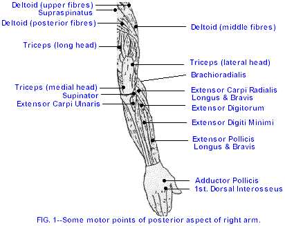
SEGMENTAL INNERVATION OF MUSCLES OF THE LOWER LIMB
TABLE I
Muscle (Segmental Innervation) Peripheral Nerve points of both heads of the gastrocnemius and of the soleus.
With a little practice, any tenderness at these motor points may be quickly elicited, although some points are
L2 Sartorius (L2, L3), Pectineus (L2. L3), Adductor longus (L.2, 1.3), L3 Quadriceps femoris (L2-L4) L4* Quadriceps femoris (L2-L4), Tensor fascise lame (L4, L5), Superior gluteal, Tibialis anterior (L4, L5), L5 Gluteus medius (L4-S1), Semimem>xanosus (L4-S1), Semitendinosus (L4-S1), Extensor hallucis longus (L4-S1) , S1 Gluteus maximus (L4-S2), Biceps femoris, short head (L5-S2), Semitendinosus (L4-S1), Medial gastrocnemius (S1, S2), Soleus (S1, S2) S2 Biceps femoris, long head (S1, S2), Lateral gastrocnemius (S1, S2), Soleus (S1, S2)
* Muscles receive innervation from more than one segment. The segments listed on the left are those generally accepted as the predominant source of innervation of the muscles in question. All are innervated by the anterior rami; the posterior rami go to corresponding levels of the erector spinae muscles, but there is extensive overlapping of the posterior rami.
In this prospective study, 100 patients and 100 control subjects were examined and followed. They were divided into four groups:
Patients with Low-Back Pain (Groups A and B)100 patients with low-back symptoms were selected from 147 consecutive patients. 47 patients were excluded with compression fractures, advanced degenerative osteoarthritic changes, and previous laminectomy or spine fusion.
All patients were managed with the standard Clinic regimen 8 including physiotherapy followed by graduated remedial exercises as well as occupational therapy or industrial activities as tolerance improved. Patients also received instruction in the care of the back and in proper bending and lifting techniques.
Control Subjects (Groups C and D)We decided to use 100 members of the lay staff of the Workers’ Compensation Board as controls. Their combined age and sex distributions were comparable to those of the patients in Groups A and B.
The control subjects were divided into two groups: Group C, fifty men and women who had no back disability; and Group D, fifty men and women who considered themselves normal, without back discomfort at the time although they had had occasional low-back discomfort after unusual activity.
Results
Group A - Low-Back SprainNo history of previous back surgery, no radicular symptoms, and no feeling of weakness, numbness, or paresthesia. No radicular signs, reflex changes, sensory changes, motor weakness, or muscle atrophy. Roentgenograms were normal or showed no more than minimum degenerative changes consistent with age or minor congenital abnormalities. No spondylolysis or spondylolisthesis. Of these fifty patients, there were twenty-six (52 per cent) who had tender motor points and twenty-four (48 per cent) who did not. These two subgroups were compared with respect to roentgenographic changes, mechanism of injury, and duration of disability. 15 of the 26 patients with tender motor points had roentgenographic abnormalities, while only two of the twenty-four without tender points had such findings.
The duration of disability ranged from 12 to 34 weeks (average, 19.7 weeks) in 25 of the 26 patients with tender motor points. In the 24 patients with no tender motor points, the disability period ranged from 3 to 13 weeks (average, 6.9 weeks).
Group B - Disc InvolvementThis Group had radicular symptoms and signs. All but one had tender motor points. The exception sustained a contusion of the hamstring muscles and this, rather than radicular involvement, was responsible for the limited straight leg raising. Although many of the patients with tender motor points had unilateral symptoms, as often as not their tender motor points were bilateral.
The duration of disability of the 49 patients in Group B with tender motor points ranged from 14 to 72 weeks (average, 25.7 weeks), while the one patient with no tender motor points was disabled for only 8 weeks.
Group C - No Back DisabilityThese subjects with no back disability showed no positive findings, developed Grade-1 tender motor points after unusual activity, such as jogging, or shoveling snow; their tenderness disappeared a few days later, only to recur whenever they increased their activities.
Group D - Occasional Back Discomfort
Physical examinations in Group-D subjects with occasional back pain were negative at the time of examination. But 46 (92%) had Grade-1 or Grade-2 tender motor points.
No correlations were evident between the locations, numbers, and grades of the tender points and the location of the degenerative changes visible on the roentgenograms.
Discussion
It is generally agreed that virtually everyone eventually has some degenerative joint disease in the low back, but that as a rule problems arise only when the degeneration has reached a certain degree and some incident, which may be minor, precipitates symptoms.
In this study it was found that an injury involving flexion combined with rotation of the lumbar spine is most likely to cause prolonged disability and that tender motor points may be useful in assessing back problems, particularly when no positive physical signs are detectable.
Tender motor points of a mild and transient nature may occasionally be found in asymptomatic individuals, especially after unusual activity. Moderately tender motor points are usually present in so-called vulnerable backs or lesser degrees of trauma. The presence of tender motor points might be significant in pre-employment medical examinations. Moderately to acutely tender motor points are almost constantly found in patients with disc degeneration. The degree of tenderness and the number of tender points tend to parallel the patient’s condition and may serve as indicators of progress.
An important finding was that low-back patients and no tender points were disabled for an average of 6.9 weeks, while those with tender points were disabled for an average of 19.7 weeks, almost as long as patients with radicular signs, who were disabled for an average of 25.7 weeks. Tender motor points, may, therefore, be a sensitive indicator of radicular involvement. Recovery time may be related to the degree of trauma sustained. Patients seen for the first time, who show no physical signs except tender motor points, deserve attention and continued surveillance.
Conclusions
Low-back pain without significant physical signs may present a diagnostic challenge. Tender motor points may be a clue under these circumstances.
This study suggests that muscle tenderness, maximum at motor points, can be elicited during the routine examination of the back and be a useful diagnostic and prognostic sign in this enigmatic group of low-back sprains.
Patients diagnosed as having simple low-back sprain but demonstrating acutely tender motor points will have a period of disability approaching that of patients with radicular signs, while patients with no tender motor points can be expected to do well.
References
1. Chusid, JG: Correlative Neuroanatomy and Functional Neurology. Ed. 15, pp. 236-237. Los Altos, California, Lange Medical Publications, 1973.2. Coers, C: Note sur une technique de prelevement des biopsies neuro-musculaires. Acta Neurol. Psychiat. Belgica, 53:759-765, 1953.3. Cyriax, JH: Textbook of Orthopaedic Medicine. Ed. 5, p. 646. London, BaiUierc, Tindall and Cassell, 1969.4. Denny-Brown, D, and Brenner, Charles: The Effect of Percussion of Nerve. J. Neurol•, Neurosurg., and Psychiat., 7: 76-95, 1944.5. Goodgold, Joseph, and Eberstein, Arthur: Electrodiagnosis of Neuromuscular Diseases, pp. 3 and 164. Baltimore, Williams and Wilkins, 1972.6. Gunn, CC, and Milbrandt, WE:Tennis Elbow and the Cervical Spine. Canadian Med. Assn, J., 114: 803-809, 1976.7. Gunn, CC, and Milbrandt, WE: Unpublished data.8. Milbrandt, WE, and Gunn, CC: A Comprehensive and Progressive Rehabilitation Programme for Low Back Strain as applied at the Workers’ Compensation Board of British Columbia. In Proceedings, International Symposium on the Rehabilitation of the Industrially Injured. Vancouver, British Columbia, Canada, April 1973.9. Nassim, Reginald and Burrows, HJ: Modern Trends in Diseases of the Vertebral Column, p. 268. London, Butterworths, 1959
IMS (intramuscular stimulation) from the Ottawa Physiotherapy and Sport Clinics
Tenderness at Motor Points(Extract from an article that was printed in the Journal of Bone and Joint Surgery September 1976)
A DIAGNOSTIC AND PROGNOSTIC AID FOR LOW-BACK INJURY
BY CC GUNN, MA, MB, B.CHIR*, AND WE MILBRANDT, MD*,VANCOUVER, BRITISH COLUMBIA, CANADA
From the Workers’ Compensation Board, Rehabilitation Clinic, Vancouver
The following extract is included because it was the first physical sign we discovered that is related to neuropathy.
ABSTRACT: In patients with low-back injury the motor points of some muscles may be tender. Of fifty patients with low-back “strain”, twenty-six had tender motor points and twenty-four did not, while forty-nine of fifty patients with radicular signs and symptoms suggesting disc involvement had tender motor points, and the one without such tender points had a hamstring contusion which limited straight leg raising. Of fifty controls with no back disability, only seven had mild tender points after strenuous activity, while forty-six of another fifty controls with occasional back discomfort had mild motor-point tenderness. In all instances the tender motor points were located in the myotomes corresponding to the probable segmental levels of spinal injury and of root involvement, when present.
Patients with low-back strain and no tender motor points were disabled for an average of 6.9 weeks, while those with the same diagnosis but tender motor points were disabled for an average of 19.7 weeks, or almost as long as the patients with signs of radicular involvement, who were disabled for an average of 25.7 weeks. Tender motor points may therefore be of diagnostic and prognostic value, serving as sensitive localizers of radicular involvement and differentiating a simple mechanical low-back strain from one with neural involvement.
It is often difficult, if not impossible, to establish the cause of disability and to assess its degree in patients with low-back pain. While in some patients the diagnosis can be made with no difficulty on the basis of the clinical history and physical examination, in others additional diagnostic tests including myelography and electromyography may be required. As a general rule, however, such tests are reserved for patients whose diagnosis is not clinically apparent or who are expected to require surgery. There remain, therefore, many patients with no localizing physical findings for whom ancillary tests are not considered necessary. The injuries in these patients are conveniently labeled “low-back sprain”.
The physician, unable to make a firm diagnosis, may rightly or wrongly relate the pain to socio-economic and psychophysiological factors, or may even suspect malingering. Therefore, many patients with genuine discomfort may not be treated appropriately simply because there are no significant physical findings.
The Workers’ Compensation Board of British Columbia operates an Outpatient Rehabilitation Clinic to provide treatment after industrial injuries. So-called low-back sprain, a vague term encompassing a multitude of disorders, is one of the most common disabilities seen at the Clinic. In 1974, the total number of admissions for all types of injuries was almost 5,000, and 1,630 (33 per cent) of these were for injuries to the lumbar spine. Of these lumbar-spine injuries, 1,401 (86%) were given a working diagnosis of low-back sprain. The remainder were fractures and postoperative conditions after laminectomies and spine fusions 8.
While performing electromyographic examinations in this Clinic, we discovered that some patients had tenderness at the motor points. Initially, these tender areas were confirmed as being located at the motor points by showing that they were at sites where the minimum electrical stimulus evoked muscle twitches using a standard calibration-stable stimulator with variable control of outputs. These studies established that the motor points of certain muscles are frequently tender in patients with low-back pain. Electromyography also showed evidence of neuropathy in the nerves supplying these tender muscles, including increased insertion activity, more polyphasic action potentials, and prolongation of the mean duration of the motor-unit action potentials, their mean amplitude remaining normal or decreasing and a partial interference pattern being obtained even during maximum voluntary effort6,7.
Tenderness parallel the severity of the symptoms and varied from week to week and even from day to day. Localized tenderness was not found in patients having hysteria or malingering. Because of these findings, we had to revise many previous diagnoses. For example, a dull ache localized to a small area in the upper lateral quadrant of the buttock, which previously had been attributed to gluteal bursitis, was found to be a tender gluteus medius. Similarly, tenderness at the gluteus maximus had been mistaken for sciatic-nerve tenderness. Tenderness described as trochanteric bursitis was found to be located at the tensor fasciae latae motor point, while tenderness thought to be caused by “adductor strain” and “rider’s sprain” was found to be located at the motor points of the pectineus and adductor longus 3. Tenderness was rarely limited to one motor point and a search for other points in muscles of the same myotome usually revealed their presence.
Methods and MaterialExamination for Tender Motor Points
Representative muscles of the second through the fifth lumbar and the first two sacral myotomes are examined. Trauma to a nerve root causes irritation or degeneration of nerve fibers, or both. These lesions may be detected during electromyography as increased insertional activity, polyphasic action potentials,
fasciculation potentials, fibrillations, and positive sharp waves, or in the procedure described here as tender motor points. Examination of the paraspinal muscles innervated by the posterior primary rami is also necessary to confirm the pathological process involves the segmental nerve at the root level.
SEGMENTAL INNERVATION OF MUSCLES OF THE LOWER LIMB
TABLE I
Muscle (Segmental Innervation) Peripheral Nerve points of both heads of the gastrocnemius and of the soleus.
With a little practice, any tenderness at these motor points may be quickly elicited, although some points are
L2 Sartorius (L2, L3), Pectineus (L2. L3), Adductor longus (L.2, 1.3), L3 Quadriceps femoris (L2-L4) L4* Quadriceps femoris (L2-L4), Tensor fascise lame (L4, L5), Superior gluteal, Tibialis anterior (L4, L5), L5 Gluteus medius (L4-S1), Semimem>xanosus (L4-S1), Semitendinosus (L4-S1), Extensor hallucis longus (L4-S1) , S1 Gluteus maximus (L4-S2), Biceps femoris, short head (L5-S2), Semitendinosus (L4-S1), Medial gastrocnemius (S1, S2), Soleus (S1, S2) S2 Biceps femoris, long head (S1, S2), Lateral gastrocnemius (S1, S2), Soleus (S1, S2)
* Muscles receive innervation from more than one segment. The segments listed on the left are those generally accepted as the predominant source of innervation of the muscles in question. All are innervated by the anterior rami; the posterior rami go to corresponding levels of the erector spinae muscles, but there is extensive overlapping of the posterior rami.
In this prospective study, 100 patients and 100 control subjects were examined and followed. They were divided into four groups:
Patients with Low-Back Pain (Groups A and B)100 patients with low-back symptoms were selected from 147 consecutive patients. 47 patients were excluded with compression fractures, advanced degenerative osteoarthritic changes, and previous laminectomy or spine fusion.
All patients were managed with the standard Clinic regimen 8 including physiotherapy followed by graduated remedial exercises as well as occupational therapy or industrial activities as tolerance improved. Patients also received instruction in the care of the back and in proper bending and lifting techniques.
Control Subjects (Groups C and D)We decided to use 100 members of the lay staff of the Workers’ Compensation Board as controls. Their combined age and sex distributions were comparable to those of the patients in Groups A and B.
The control subjects were divided into two groups: Group C, fifty men and women who had no back disability; and Group D, fifty men and women who considered themselves normal, without back discomfort at the time although they had had occasional low-back discomfort after unusual activity.
Results
Group A - Low-Back SprainNo history of previous back surgery, no radicular symptoms, and no feeling of weakness, numbness, or paresthesia. No radicular signs, reflex changes, sensory changes, motor weakness, or muscle atrophy. Roentgenograms were normal or showed no more than minimum degenerative changes consistent with age or minor congenital abnormalities. No spondylolysis or spondylolisthesis. Of these fifty patients, there were twenty-six (52 per cent) who had tender motor points and twenty-four (48 per cent) who did not. These two subgroups were compared with respect to roentgenographic changes, mechanism of injury, and duration of disability. 15 of the 26 patients with tender motor points had roentgenographic abnormalities, while only two of the twenty-four without tender points had such findings.
The duration of disability ranged from 12 to 34 weeks (average, 19.7 weeks) in 25 of the 26 patients with tender motor points. In the 24 patients with no tender motor points, the disability period ranged from 3 to 13 weeks (average, 6.9 weeks).
Group B - Disc InvolvementThis Group had radicular symptoms and signs. All but one had tender motor points. The exception sustained a contusion of the hamstring muscles and this, rather than radicular involvement, was responsible for the limited straight leg raising. Although many of the patients with tender motor points had unilateral symptoms, as often as not their tender motor points were bilateral.
The duration of disability of the 49 patients in Group B with tender motor points ranged from 14 to 72 weeks (average, 25.7 weeks), while the one patient with no tender motor points was disabled for only 8 weeks.
Group C - No Back DisabilityThese subjects with no back disability showed no positive findings, developed Grade-1 tender motor points after unusual activity, such as jogging, or shoveling snow; their tenderness disappeared a few days later, only to recur whenever they increased their activities.
Group D - Occasional Back Discomfort
Physical examinations in Group-D subjects with occasional back pain were negative at the time of examination. But 46 (92%) had Grade-1 or Grade-2 tender motor points.
No correlations were evident between the locations, numbers, and grades of the tender points and the location of the degenerative changes visible on the roentgenograms.
Discussion
It is generally agreed that virtually everyone eventually has some degenerative joint disease in the low back, but that as a rule problems arise only when the degeneration has reached a certain degree and some incident, which may be minor, precipitates symptoms.
In this study it was found that an injury involving flexion combined with rotation of the lumbar spine is most likely to cause prolonged disability and that tender motor points may be useful in assessing back problems, particularly when no positive physical signs are detectable.
Tender motor points of a mild and transient nature may occasionally be found in asymptomatic individuals, especially after unusual activity. Moderately tender motor points are usually present in so-called vulnerable backs or lesser degrees of trauma. The presence of tender motor points might be significant in pre-employment medical examinations. Moderately to acutely tender motor points are almost constantly found in patients with disc degeneration. The degree of tenderness and the number of tender points tend to parallel the patient’s condition and may serve as indicators of progress.
An important finding was that low-back patients and no tender points were disabled for an average of 6.9 weeks, while those with tender points were disabled for an average of 19.7 weeks, almost as long as patients with radicular signs, who were disabled for an average of 25.7 weeks. Tender motor points, may, therefore, be a sensitive indicator of radicular involvement. Recovery time may be related to the degree of trauma sustained. Patients seen for the first time, who show no physical signs except tender motor points, deserve attention and continued surveillance.
Conclusions
Low-back pain without significant physical signs may present a diagnostic challenge. Tender motor points may be a clue under these circumstances.
This study suggests that muscle tenderness, maximum at motor points, can be elicited during the routine examination of the back and be a useful diagnostic and prognostic sign in this enigmatic group of low-back sprains.
Patients diagnosed as having simple low-back sprain but demonstrating acutely tender motor points will have a period of disability approaching that of patients with radicular signs, while patients with no tender motor points can be expected to do well.
References
1. Chusid, JG: Correlative Neuroanatomy and Functional Neurology. Ed. 15, pp. 236-237. Los Altos, California, Lange Medical Publications, 1973.2. Coers, C: Note sur une technique de prelevement des biopsies neuro-musculaires. Acta Neurol. Psychiat. Belgica, 53:759-765, 1953.3. Cyriax, JH: Textbook of Orthopaedic Medicine. Ed. 5, p. 646. London, BaiUierc, Tindall and Cassell, 1969.4. Denny-Brown, D, and Brenner, Charles: The Effect of Percussion of Nerve. J. Neurol•, Neurosurg., and Psychiat., 7: 76-95, 1944.5. Goodgold, Joseph, and Eberstein, Arthur: Electrodiagnosis of Neuromuscular Diseases, pp. 3 and 164. Baltimore, Williams and Wilkins, 1972.6. Gunn, CC, and Milbrandt, WE:Tennis Elbow and the Cervical Spine. Canadian Med. Assn, J., 114: 803-809, 1976.7. Gunn, CC, and Milbrandt, WE: Unpublished data.8. Milbrandt, WE, and Gunn, CC: A Comprehensive and Progressive Rehabilitation Programme for Low Back Strain as applied at the Workers’ Compensation Board of British Columbia. In Proceedings, International Symposium on the Rehabilitation of the Industrially Injured. Vancouver, British Columbia, Canada, April 1973.9. Nassim, Reginald and Burrows, HJ: Modern Trends in Diseases of the Vertebral Column, p. 268. London, Butterworths, 1959
IMS (intramuscular stimulation) from the Ottawa Physiotherapy and Sport Clinics
 In the morning, before breakfast when your blood sugar levels are low and it is ideal for fat burning exercises ,you will use up energy for exercising from fat stored in your body instead of energy from carbohydrates you have just eaten. If you exercise immediately after breakfast you will still burn the fat but only after you burn the calories from breakfast first. You can burn as much as 3 times more fat when cardio exercises are done on an empty stomach.